Click here for a full equipment list across all School of Medicine Research Cores
Microscopes
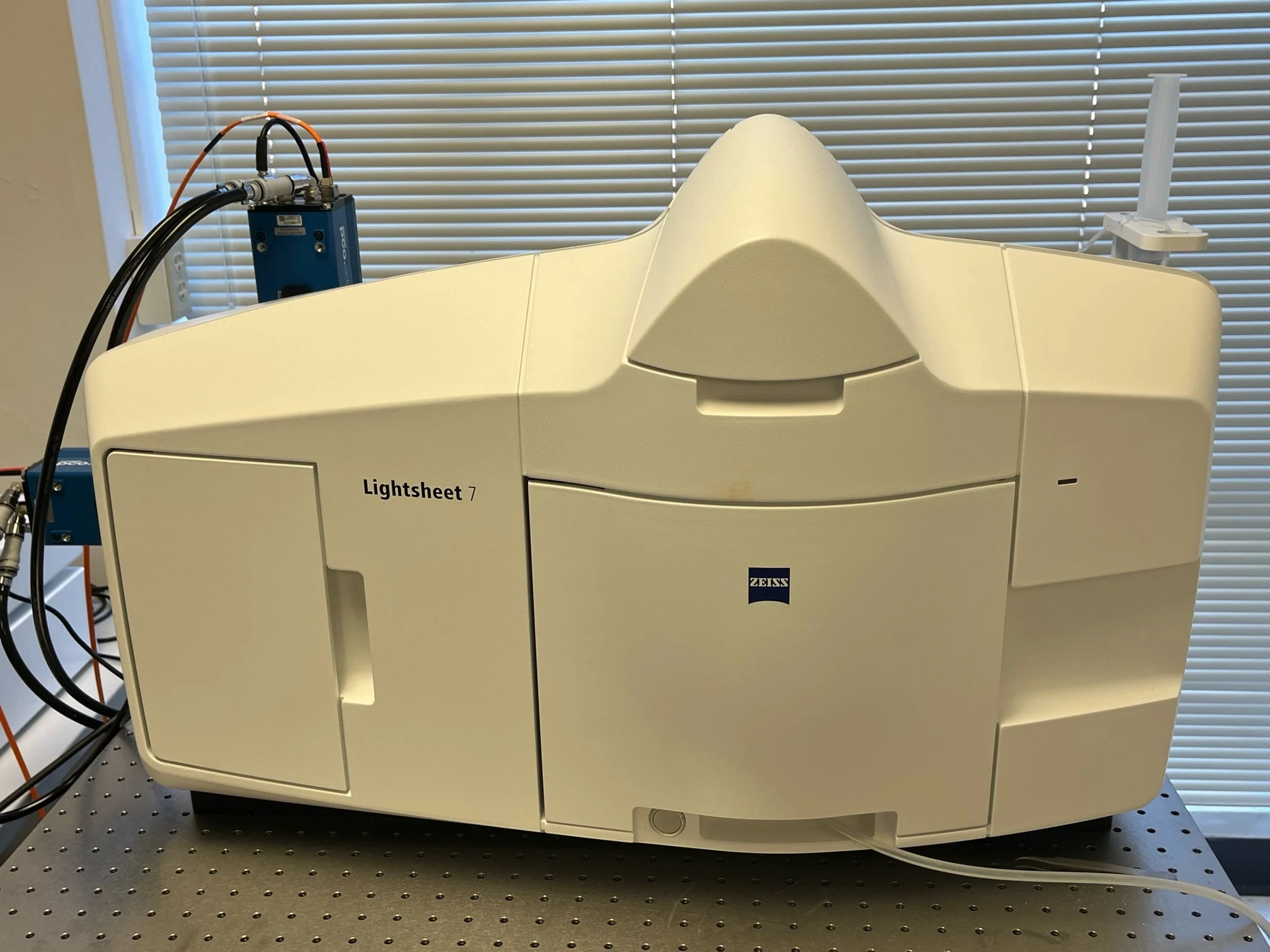
Zeiss Lightsheet 7
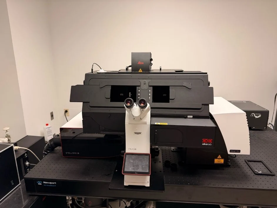
Standalone sealed box system (laser safe) for multiview imaging of living and optically cleared specimens up to 2cm in size at subcellular resolution given any refractive index between 1.33 – 1.58
Inverted spectral imaging confocal microscope equipped with AiryScan 2 which enables 2x higher resolution images at high speed acquisition, and an ensemble of 2 multi-alkaline PMTs, 32-channel spectral GaAsP, and 2 near-IR GaAs detectors
Equipped with Ai sample finder for automatically locating tissue section
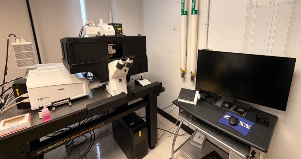
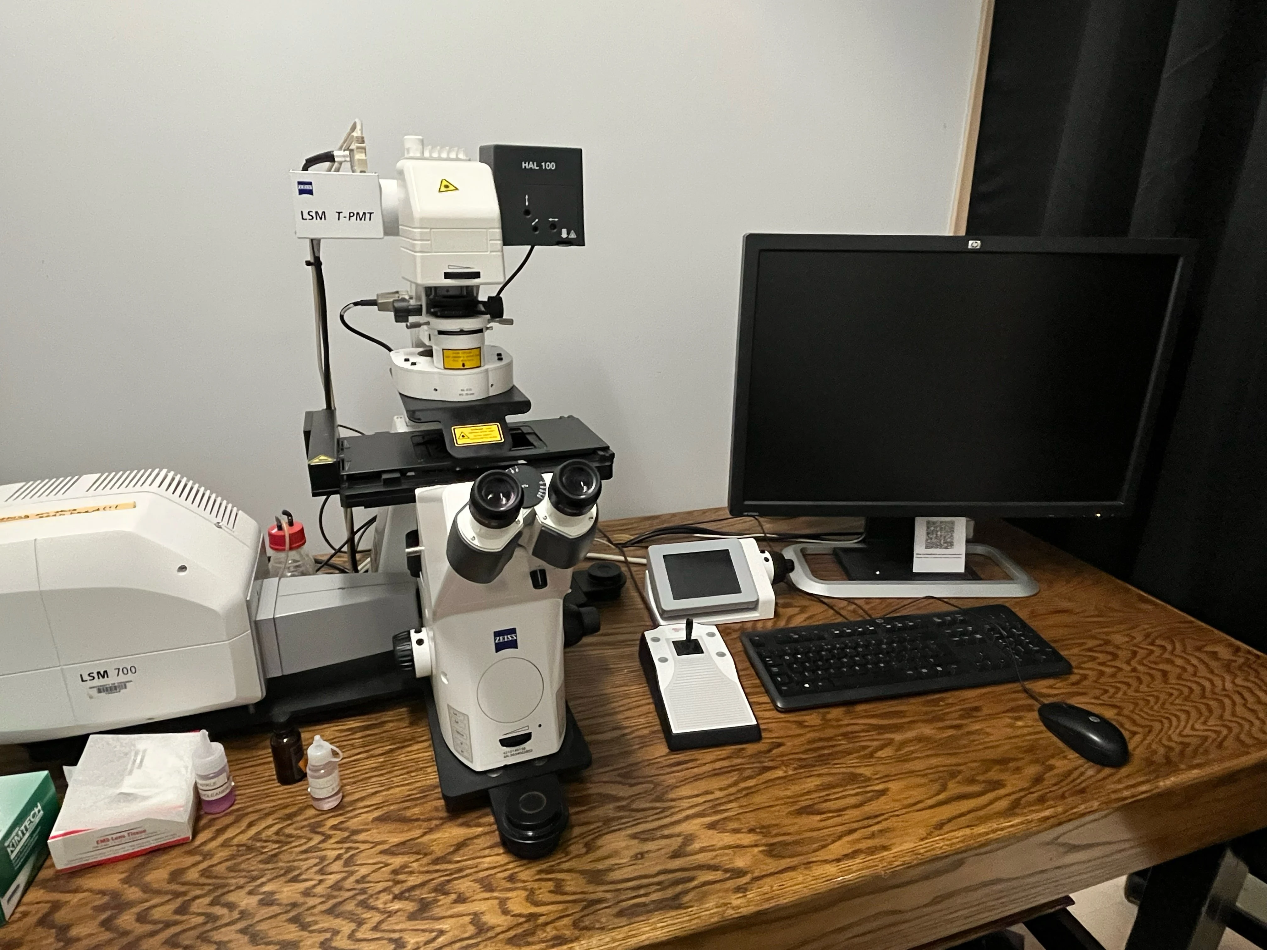
Inverted spectral imaging confocal microscope equipped with AiryScan/fastAiryScan which enables 1.7x higher resolution images at high speed acquisition
Equipped with a 34 channel spectral array
Laser lines: 405nm, 458nm, 488nm, 514nm, 543nm, 594nm and 633nm
Objectives: 10x, 20x, 40x, and 63x
Long-term live cell imaging: includes a fully enclosed CO2 chamber
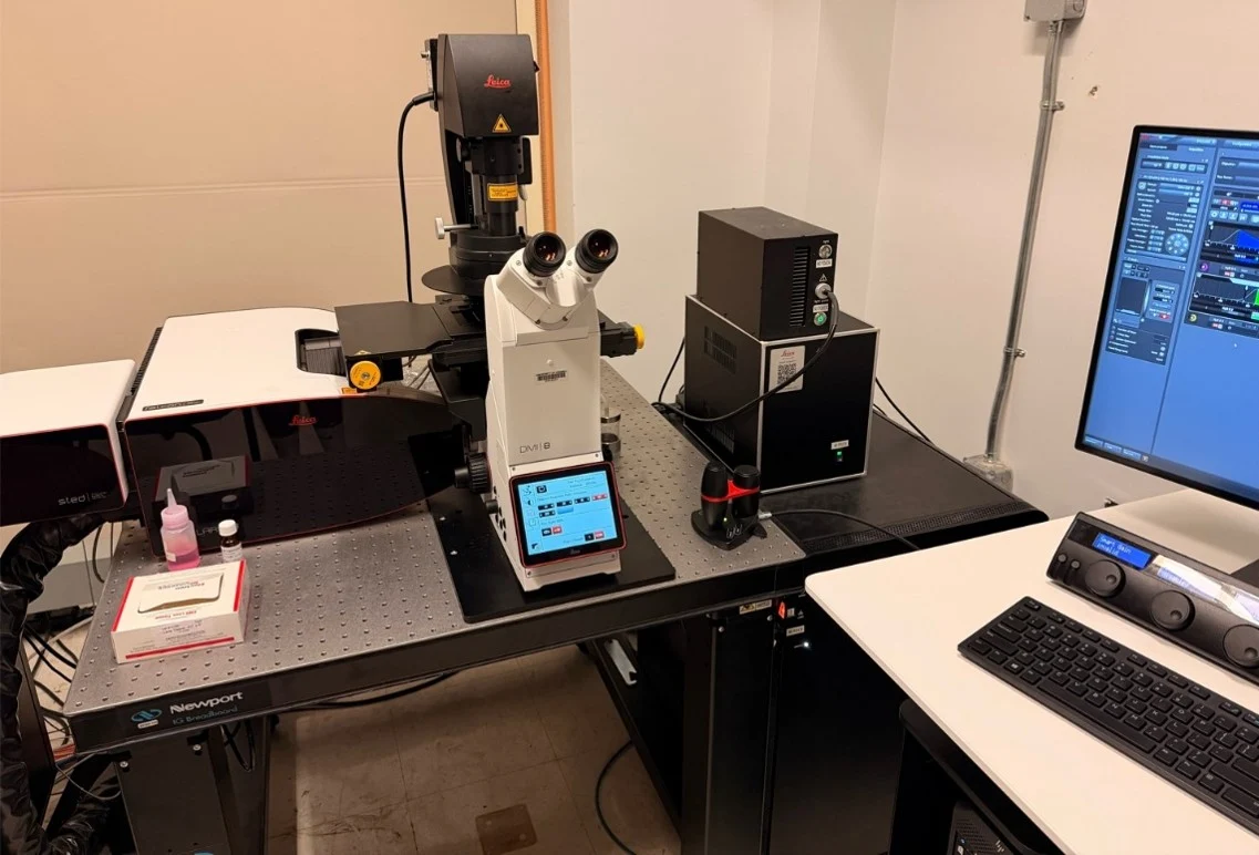
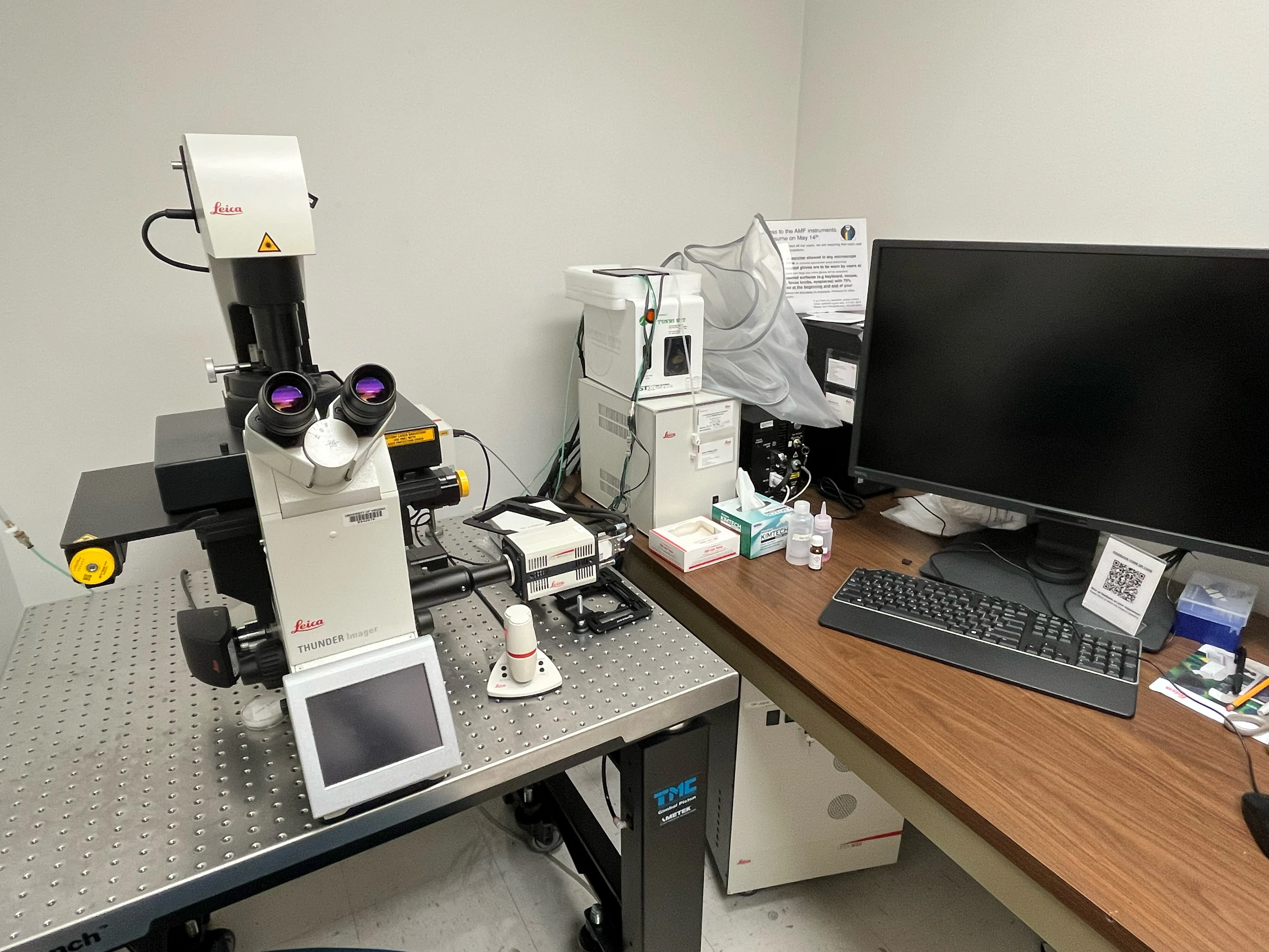
Fully automated inverted microscope equipped with a W-View Gemini for dual color TIRF as well as color and monochromatic cameras for widefield and brightfield, respectively
Laser lines for TIRF: 405, 488, 561, and 638nm
Objectives: 5x, 10x, 20x, 40x, 63x
Live cell imaging: includes CO2 connection with stage top incubation
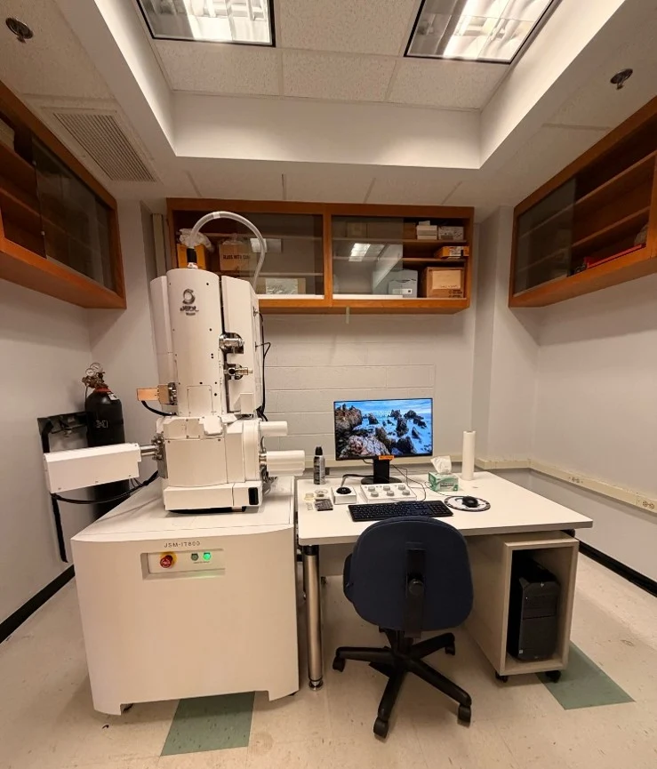
The integrated In-Lens Schottky Plus and low aberration condenser lens achieves high beam brightness. Ample probe current (100nA@5kV) is available even at low accelerating voltage.
Stable observation is possible with Neo Engine (New Electron Optical Engine) while adjusting various microscope parameters.
The hybrid lens (HL) lens are equipped with an electromagnetic/electrostatic field superposed objective lens developed from the versatile out-lens. The high resolution observation and analysis of a wide variety of specimens from metals to nano materials are possible.
Scintillator Backscattered Electron Detector (SBED)