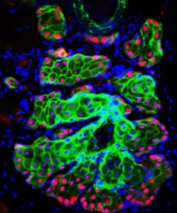
Fluorescence image showing the Meibomian gland stained for K14 (green), an epithelial cell marker, and PPARg, which labels meibocytes, the cells that produce meibum and originate from Krox20+ stem cells. DAPI (blue) stains nuclei.
Dry Eye Disease is a common ophthalamic problem and 90% of cases are caused by Meibomian Gland Dysfunction. The Meibomian glands are holocrine sebaceous glands located in the tarsal plates of the upper and lower eyelids. They contain meibocytes, epithelial cells that rupture and release their lipid content called meibum, which maintains moisture on the ocular surface by protecting the aqueous layer from evaporation. The stem cells that give rise to and maintain homeostasis of this gland remain elusive. Studies by Tchegnon et al. revealed that loss of Krox20, a zinc-finger transcription factor, disrupts Meibomian gland formtion and homeostasis, leading to Dry Eye Disease secondary to Meibomian Gland Dysfunction.
Read the full publication here.

Comments