Services
Click here for a full service list across all School of Medicine Research Cores
Primary Techniques
The primary techniques used here are:
- unstained, unfixed samples
- single particles from solutions with potential resolution < 1nm
- helical and two-dimensional crystals
- some atomic models rival X-ray crystallography and NMR
- 3-D maps of non-regular structures, larger cellular complexes and cell sections
- uses tilt series to generate 3-D structure
- resolution 3-4 nm for irregular structures, higher for regular structures
- electron diffraction of micro-crystals
- uses tilt series to generate diffraction patterns
- resolution can reach 1-2Å
- guidance and suggestions for data processing
- direct support in setting up appropriate servers or workstations
- limited data analysis for single-particle and tomographic projects
- provide limited Molecular Dynamics simulations
- provide searches of the full AlphaFold database
Using the facilities
 Trained researchers can use microscopes themselves. Alternatively, MEMC personnel can assist researchers in collecting images.
Please contact Michael Purdy (mpurdy@virginia.edu) to discuss how best to use the facility to aid your project. A project application will be required, at which time access to the scheduling system will be provided.
Clients will need to provide a 8-12Tb USB storage drive for multi-day data collection on the Titan Krios or download data from MEMC servers. Storage of data will only be guaranteed for one month. Images collected on the Spirit or F20 will accessible by download from a remote server.
Sample preparation
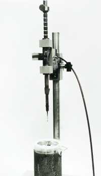
Researchers can freeze samples in liquid ethane with a manual guillotine or using an FEI Vitrobot. Several options are also available for negative-staining.
Typical sample requirements pre grid are 3 µL at concentrations of 0.1-1.0 mg/ml. A minimum of 20 µL is preferred in order to freeze multiple grids.
Frozen Sample
By examining frozen, unstained samples in these high resolution transmission electron microscopes and taking images from different angles, researchers get structural data down to the molecular level, without having to crystallize proteins. This data complements the expertise in protein X-ray crystallography and NMR in the Department of Molecular Physiology and Biophysics
