Equipment
Click here for a full equipment list across all School of Medicine Research Cores
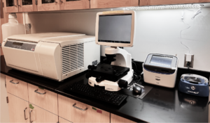
The nCounter Pro Analysis System and the Sprint Profiler provide a cost-effective automated solution for multiplex analysis of up to 800 targets. The simple workflow requires just 15 minutes of hands-on time and produces highly reproducible data, requiring no amplification or technical replicates.
The panels cover different areas of research, such as Oncology, Immunology, Cardiovascular Diseases, Cell & Gene Therapy, Stem Cells, Regenerative Medicine, Infections Disease, Neuroscience, etc.
Customizing panels is another available tool. It is possible to include different targets in the chosen panel or customize your panel.
nCounter can be a shift option in studies that use the PCR technique, eliminating primer design and validation steps and optimizing the use of biological samples without technical replicates.
The nCounter technology requires no amplification or cDNA synthesis.
Analyzes can be performed using nSolver or Rosalinda software, both online platforms. Both software is easy to use, generating high throughput data and publishable pictures in less than 1 hour.
More information:
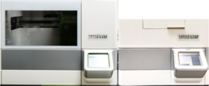
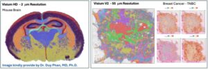
The Visium CytAssist is an instrument designed to transfer transcriptomic probes from standard glass slides to Visium slides.
Whole transcriptome spatial discovery in a single-cell scale.
Visium HD Spatial Gene Expression slides contain two 6.5 x 6.5 mm Capture Areas with a continuous lawn of oligonucleotides arrayed in ~11 million 2 x 2 μm barcoded squares without gaps, achieving single–cell–scale spatial resolution.
More information:
If you have an interest or want to know more about this platform, please contact Dr Ana de Oliveira at ak4yj@virginia.edu
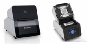
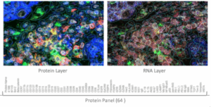
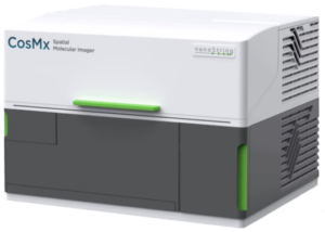
CosMxTM Spatial Molecular Imager (SMI) is the first high-plex in situ analysis platform to provide spatial multi-omics with formalin-fixed paraffin-embedded (FFPE) and fresh frozen (FF) tissue samples at cellular and subcellular resolution. CosMx SMI enables rapid quantification and visualization of 1,000 or 6,000 RNA and 64 or 120 proteins at the same time. It is the flexible, spatial single-cell imaging platform that will drive deeper insights for cell atlasing, tissue phenotyping, cell-cell interactions, cellular processes, and biomarker discovery.
The CosMxTM SMI platform is an integrated system with mature cyclic in situ hybridization chemistry, an ultra-high-resolution imaging readout instrument, and interactive data analysis and visualization software.
CosMx Same-Cell MultiOmics
Performing spatially-resolved multi-omics analysis on the same tissue section.
Run the RNA and Protein panels in the same tissue using CosMx platform. This workflow enable the detection of over 19,000, 6,000 or 1,000 RNA and 64 proteins (+ 8 custom protein) in the FFPE samples.
Applications:
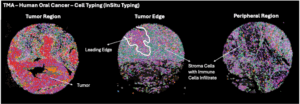
- Cell phenotyping
- Tissue context (cells vs stroma structures)
- Validate the candidate biomarkers directly in tissue by aligning protein detection with RNA expression at subcellular resolution
More information:
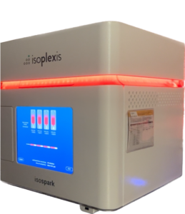
IsoCode Chip – Single-Cell Analysis
Isoplexis is a multiplex phenotyping analysis that allows complete functional characterization of a single-cell.
Cells are initially stained (2 markers) and captured individually in the chip’s microchambers. Over 30 cytokines, chemokines, and a full range of phosphoproteins are monitored for 20 hours.
Panel List:
Single-Cell Secretome
Human Adaptive Immune
Granzyme B, IFN-γ, MIP-1α, Perforin, TNF-α, TNF-β, GM-CSF, IL-2, IL-5, IL-7, IL-8, IL-9, IL-12, IL-15, IL-21, CCL11, IP-10, MIP-1β, RANTES, IL-4, IL-10, IL-13, IL-22, TGFβ1, sCD137, sCD40L, IL-1β, IL-6, IL-17A, IL-17F, MCP-1, MCP-4
Human Natural Killer
Granzyme B, IFN-γ, MIP-1α, Perforin, TNF-α, TNF-β, GM-CSF, IL-2, IL-5, IL-7, IL-8, IL-9, IL-12, IL-15, IL-21, CCL11, IP-10, MIP-1β, RANTES, IL-4, IL-10, IL-13, IL-22, TGFβ1, sCD137, sCD40L, IL-1β, IL-6, IL-17A, IL-17F, MCP-1, MCP-4
Human Innate Immune
IFN-γ, MIP-1α, TNF-α, TNF-β, GM-CSF, IL-8, IL-9, IL-15, IL-18, TGF-α, IL-5, CCL11, IP-10, MIP-1β, RANTES, BCA-1, IL-10, IL-13, IL-22, sCD40L, IL-1β, IL-6, IL-12-p40, IL-12, IL-17A, IL-17F, MCP-1, MCP-4, MIF, EGF, PDGF-BB, VEGF
Human Inflammation
GM-CSF, IFN-γ, IL-2, IL-12, TNF-α, TNF-β, IL-4, IL-5, IL-7, IL-9, IL-13, CCL11, IL-8, IP-10, MCP-1, MCP-4, MIP-1α, MIP-1β, RANTES, IL-10, IL-15, IL-22, TGF-β1, IL-1β, IL-6, IL-17A, IL-17F, IL-21, Granzyme B, Perforin, sCD40L, sCD137
Mouse Adaptive Immune
Granzyme B, IFN-γ, MIP-1α, TNF-α, GM-CSF, IL-2, IL-5, IL-7, IL-12p70, IL-15, IL-21, sCD137, CCL11, CXCL1, CXCL13, IP-10, RANTES, Fas, IL-4, IL-10, IL-13, IL-27, TGFβ1, IL-6, IL-17A, MCP-1, IL-1β
Single-Cell Signaling
Human Adaptive Immune
TNF-a, IFN-g, PERFORIN, GRANZYME B, IL-10, MIP-1b, IL-2, GM-CSF, P-IkBA, P-NF-kB p65, P-Stat3, P-MEK1/2, P-Stat1, P-Stat5, IL-8
CodePlex Chip – Secretome Analysis in Supernatant or Biological Fluid
- Completely automated workflow
- 5.5 uL per sample in duplicate
- Measure more than 30 cytokines in bulk
- Run 8 samples per Chip
- Modular, load 8-32 samples per run
- Limit of detection: 5-5000 pg/ml
Panel List
Human Adaptive Immune
GM-CSF, Granzyme B, IFN-γ, IL-2, IL-4, IL-5, IL-6, IL-7, IL-8, IL-9, IL-10, IL-13, IL-15, IL-17A, IP-10, MCP-1, MIP-1α, MIP-1β, Perforin, sCD137, TNF-α, TNF-β
Human Innate Immune
EGF, GM-CSF, Granzyme B, IFN-γ, IL-1β, IL-4, IL-6, IL-7, IL-8, IL-10, IL-15, IP-10, MCP-1, MIP-1α, MIP-1β, PDGF-BB, sCD137, TNF-α, VEGF
Mouse Adaptive Immune
GM-CSF, IFN-γ, IL-1β, IL-2, IL-4, IL-5, IL-6, IL-10, IL-12, IL-17A, IP-10, KC, MCP-1, MIP-1α, RANTES, TNF-α
Mouse Innate Immune
IFN-γ, TNF-α, MIP-1α, IL-15, GM-CSF, IL-5, IL-10, IL-13, IL-6, IL-17A, MCP-1, IP-10, MIP-1β, EGF, PDGF-BB, MIF
Mouse Inflammation
IFN-γ, TNF-α, MIP-1α, IL-2, IL-5, IL-10, IL-13, IL-4, IL-6, IL-1β, IL-17A, IL-12, MCP-1, IP-10, KC, GM-CSF
Meteor
- Fully quantitative bulk analysis
Panel List
Human Adaptive Immune 1
GM-CSF, IFN-g, IL-2, IL-4, IL-5, IL-6, IL-8, IL-10, IL-17A, TNF-a
With a sample volume of just 7.5uL per sample and the capacity to run 20 samples in triplicate on each chip + calibration curve for each cytokine.
More information:
Bright field and Immunofluorescence
Magnification: 5x, 20x, 40x
Filters: DAPI, GFP, Cy3, Cy5
Software: Zeiss Zen
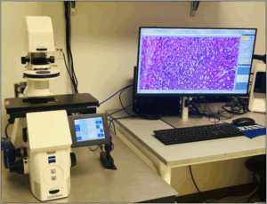
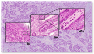
Image analysis performed by Natalia Dworak at the Spatial Biology Core. Colon Cancer.
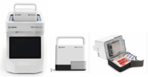
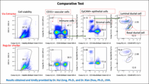
The VIA Extractor is a tissue disaggregator that processes solid tissue into single cells with high viability and yield.
More Information
The Stem Cell Lab is available again
Facilities:
Appropriate facilities for the development of iPSC and organoid culture.
All instrumentation available:
- 02 Hoods in the regular lab room
- 01 Hood in the quarantine room
- HERA Cell Vios 160i CO2 Incubator (04 incubators)
- EVOS XL Core microscope inside the hood (magnification 4x, 10x, 20x, 40x)
- EVOS FLc with for color filters (Cy5, GFP, DAPI, TxRed) and 4 magnifications
- Cell counting
- Water bath
- Centrifuge
- Sonic Dismembrator Model 500 – Fisher Scientific
- Refrigerator, -20ºC Freezer and -80ºC Freezer
Scientific support
Training support
Cell maintenance
Mycoplasma control (screening with Lonza kit)