Equipment
Click here for a full equipment list across all School of Medicine Research Cores
Electron Microscopes
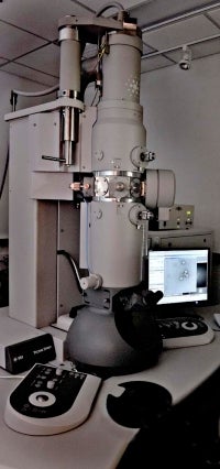 The 120kV Tecnai Spirit is equipped with a tungsten filament electron source, and a 2kx2k UltraScan CCD camera. It is primarily available for screening negative-stained samples, but is capable of cryo-electron microscopy.
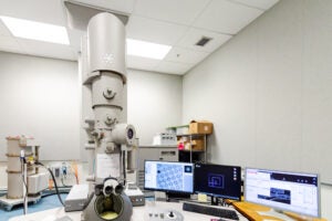 The F20 can operate at 120kV or 200kV. It has a field emission gun (FEG) source of electrons, which provides better coherence. It is used for basic electron cryomicroscopy of moderate resolution projects, or for higher-resolution negative-stain data collection. It is equipped with a 4k x 4k UltraScan CCD camera. Funded by National Institutes of Health Shared Instrumentation Grant refer to How To Acknowledge Core
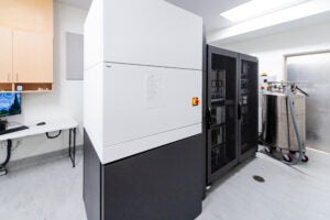
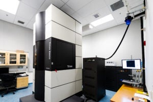 The Titan Krios has an XFEG electron source and operates at 300kV.
This microscope not only has a Falcon IIIEC direct electron detector camera, but a K3/GIF has recently been added, which avoids loss of data quality which can occur with other detectors and is more sensitive. This detector effectively increases resolution by recording sharper images and compensating for sample movement while it is imaged.
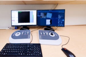
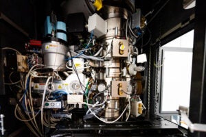 It has been designed to be extremely stable by environmentally isolating the system from external vibrations and temperature changes. Thus grids are robotically transferred into the beam and the user operates the instrument from a separate room.
The improved system provides superior controllability and reproducibility, and the ability to collect data on a single grid for up to a week. Traditionally, EM grids have to be changed manually every 4-6 hours.
Up to ten sample grids can be loaded.
For suitable specimens, computational analysis of images from the Krios with a direct detector generates atomic models that rival those calculated by X-ray crystallography and NMR spectroscopy. Funded by National Institutes of Health Shared Instrumentation Grant refer to How To Acknowledge Core
This instrument determined the structure of a highly stable virus, as published in Science
The Glacios is a 200kV electron microscope with a cryo Autoloader, XFEG electron source and Falcon4 direct electron detector. It is ideal for screening cryo samples and preliminary data collection for cryoEM and cryoET.
Sample Preparation Equipment
The GP2 provides excellent control over the process of vitrifying electron microscopy grids. In addition to maintaining the temperature and humidity of the environmental chamber, the GP2 can modulate the temperature of the liquid ethane. It has been reported that this can reduce initial beam-induced motion. The GP2 blots from one side of the grid.
The FEI Vitrobot Mark IV System allows for semi-automated vitrification to provide fast, easy, and reproducible sample preparation for cryo-EM. It provides humidity and temperature control in the sample chamber. It blots grids from both sides, which provides a gradient of ice thicknesses. It also allows users to specify the blotting times and force.
The Leica EM UC7 Ultramicrotome can produce ultra- or semi-thin sections for TEM or ultramicrotomy. The Ultramicrotome is operated by our in-house expert by appointment.
The Thermo Scientific Aquilos 2 Cryo-FIB incorporates a gallium-focused ion beam for lamella milling to produce samples as thin as 100-200 nm. This allows for tomography of biomolecules in their cellular context. The Aquilos combines light and electron microscopy in one device, allowing direct correlation of the images.
The MEMC has two Pelco easiGlow Glow Discharge systems and a Safematic CCU-010 HV high compact vacuum coating system. The lab also has various other laboratory equipment you might need to prepare samples.
