
Pathogenesis of Disc Degeneration
In the end stage of intervertebral disc degeneration, cartilage, bone, endothelial cells, and neurons appear in association with the worsening condition. The source of these different cell types in the degenerated intervertebral disc (IVD) is controversial.
One hypothesis suggests that foreign cell types migrate into the degenerating IVD from surrounding spinal tissue, while another promotes the notion that the disc contains progenitor cells that differentiate into these different cell types. Using a rabbit disc degeneration model, we previously demonstrated that injection of a 30 kDa fragment of fibronectin into the annulus fibrosus (AF) led to the formation of osteophytes in the anterior region of the AF, a peripheral region of cartilaginous tissue with a central region of osteoid and cancellous bone.
We hypothesize that the ectopic bone formation, innervation, and vascularization in the degenerative IVD is also a result of progenitor cell differentiation in the inner AF tissue because biomechanical and/or biochemical alterations change the AF progenitor cell niche, leading to differentiation of indwelling cells and disc degeneration.

Normal annulus fibrosus tissue contains multipotent progenitor cells, which are able to differentiate into cartilage and/or fibrocartilage cells, osteoblasts, neurons, and blood vessel cells.
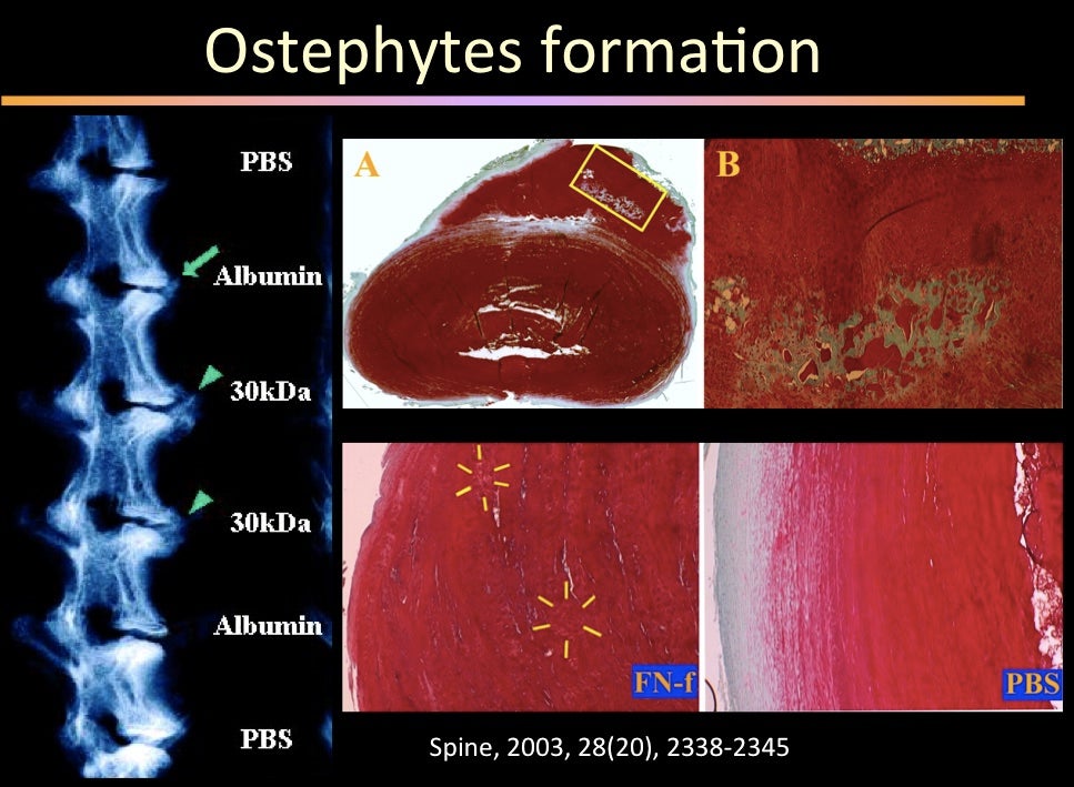
Big disc osteophyte formation following disc injury
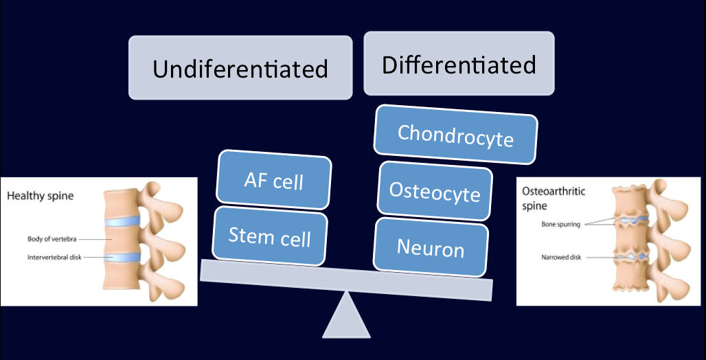
Imbalanced differentiation of disc cells in response to deterioration of local biomechanics and biochemical environment