
Procedure details
Preparation
You will first meet with a doctor who specializes in treating cancer with radiation (radiation oncologist). You may also have to undergo scans to help your doctor determine the treatment plan. Procedures such MRI, CT scans, PET scans, or x-rays may be performed prior to treatment.
Prior to your procedure you will be given specific instructions on how to prepare for your brachytherapy procedure when you meet with your radiation oncologist. Often times you will be asked to take specific medications prior to your procedure. These will depend on which procedure is being performed, but often include antibiotics and or bowel preparation. If you are having anesthesia, you will be asked to not eat anything for a certain amount of time prior to your procedure; details will be given to you prior to your treatment. You also may be asked to have a friend or family member with you on the day of your procedure, as you will need help getting home, if you are having anesthesia.
The Imageguided Brachytherapy Suite
The Image-Guided Brachytherapy Suite in the Emily Couric Clinical Cancer Center is a world-class facility, in which all aspects of the brachytherapy treatment can be provided. The suite consists of a dedicated treatment room with a CT-on-rails along with full anesthesia capabilities and a 3 bed recovery area. Because the equipment is dedicated for brachytherapy treatments, the patient does not need to be moved from the suite to other areas of the hospital for imaging, treatment, or recovery.
-
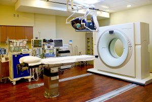
- Brachytherapy Suite: CT-on-Rails
-

- Brachytherapy Suite: CT-on-Rails
-

- Brachytherapy Suite: Recovery Area
Photo credit: Lydia Levinson
What to expect during your treatment procedure
Brachytherapy treatment involves inserting radioactive material into your body near the cancer. How the radioactive material is placed depends on the location and extent of your cancer.
Placement may be inside a body cavity or in body tissue:
- If placed inside a body cavity, a device containing radioactive material is placed in a body opening such as the vagina. The device may be a tube or cylinder made to fit that specific body opening. Imaging equipment such as a CT scanner or ultrasound machine may be used to ensure the device is placed in the correct position. For convenience, UVA has an in-room CT scanner so that the patient does not have to be moved to a separate location for imaging. Additionally, full anesthesia is available for procedures if necessary.
- If inserted into body tissue, small needles are temporarily placed to deliver radioactive material within close proximity of the radiation target. This is known as interstitial brachytherapy.
- In some instances, brachytherapy devices may be placed on the skin surface to treat superficial cancers.
High- dose rate vs low-dose rate Brachytherapy
Radiation can be given in a brief treatment session as with high-dose rate brachytherapy, or it can be left in place over a period of time as with low-dose rate brachytherapy. Sometimes the radiation source is placed in your body permanently. Again, this depends on your specific treatment.
- High-dose rate brachytherapy is typically an outpatient procedure although it will depend on your specific treatment plan. During this procedure, radioactive material is placed in your body for a short period of time (15-45 minutes). You will undergo a set number of sessions determined by your physician.
- Low-dose rate radiation is a continuous low dose of radiation released over time, typically achieved by leaving seeds in place which are positioned at the time of the brachytherapy procedure. These seeds are permanently left in the tissue but their radioactivity will decrease over time, eventually to almost no radioactivity.