
Facilities and Equipment
The Radiological Research Laboratory
The Radiological Research Laboratory occupies more than 17,000 square feet spread over three floors of the Snyder Translational Research Building in Fontaine Research Park at the University of Virginia. Additional research space and equipment is located at the main Health System campus, including a third whole-body MRI scanner, sited immediately adjacent to University Hospital.
MRI Scanners
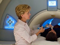
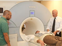 There are three MRI scanners in the UVA Main Hospital as described below. The UVA Imaging Core has 20 hours per week of dedicated research scan time on these scanners each week.
3T Siemens MAGNETOM Prisma. Features include: an actively-shielded 60cm bore, TIM [204×64] digital RF system with 64 high-speed receivers, XR Gradients (Maximum Gradient Strength 80mT/m, Maximum Slew Rate 200 T/m/s), multi-RF-coil body arrays, wireless physiological monitoring and gating system (ECG, respiratory, and pulse), advanced cardiac imaging package, cardiac DOT engine, MYOMAPS, and IDEA pulse sequence development environment.
3T Siemens MAGNETOM Skyra. Features include: an actively-shielded 70cm bore, TIM [204×64] digital RF system with 64 high-speed receivers, XQ Gradients (Maximum Gradient Strength 45mT/m, Maximum Slew Rate 200 T/m/s), multi-RF-coil body arrays, wireless physiological monitoring and gating system (ECG, respiratory, and pulse), advanced cardiac imaging package, cardiac DOT engine, MYOMAPS and IDEA pulse sequence development environment.
1.5T Siemens MAGNETOM Sola.
The Sola 1.5T offers a 70cm bore with gradient strength of 33mT/m, and slew rate of 125T/m/s. It can use up to 204 channels with the Tim 4G technology and includes quiet suite technology for patient comfort.
 Additional research scanners are available in the Snyder Building (480) that houses Radiology Research Division at Fontaine Research Park, 2 miles from the UVA Main Hospital, fully committed to clinical research.
3T Siemens MAGNETOM Prisma. Features include: an actively-shielded 60cm bore, TIM [204×64] digital RF system with 64 high-speed receivers, XR Gradients (Maximum Gradient Strength 80mT/m, Maximum Slew Rate 200 T/m/s), multi-RF-coil body arrays, wireless physiological monitoring and gating system (ECG, respiratory, and pulse), advanced cardiac imaging package, cardiac DOT engine, MYOMAPS, and IDEA pulse sequence development environment.
Avotec F-MRI equipment is available upon request.
3T Siemens CIMA MRI. 1. Cima.X is a 3T MRI system that provides deeper insights into the human body. Transforming patient care and science, it features Gemini Gradients with 2001 mT/m at 200 T/m/s for an unparalleled whole-body performance and a 60cm bore.
Siemens Biograph mCT (PETCT). Features include: 0.3 s rotation time Biograph mCT•X (0.33 s Biograph mCT•S), Biograph mCT•S: 20-, 40-, 64-slice, Biograph mCT•X: 128-slice, 78 cm open bore, Multislice UFC ™ (Ultra Fast Ceramic) detector, 100 kW generator Biograph mCT•X (80 kW Biograph mCT•S), Adaptive dose shield, Multi-LSO-detector ring system, OptisoHD detection system, 16.4 cm axial field-of-view (FoV), 22.1 cm axial FoV with TrueV, 3D acquisition mode.
A physiological monitoring and gating system (In Vivo Medical Systems, Orlando, FL) with ECG, blood pressure and pulse oximetry is available in the MR suite and a Spectris (Medrad, Indianola PA) contrast power injector is available for use.
Facilities and Other Equipment
 A 230 sq. ft. patient examination room for consent and preparation of study participants, located next to the MRI suites. Major equipment includes a handheld Koko spirometer (PDS Ferraris, Louisville, CO), a 12-lead ECG system (HP Pagewriter XLi, Hewlett Packard Co., Palo Alto, CA), and an MR compatible system for monitoring heart rate and oxygen saturation level (model 3150 MRI Patient Monitor, Invivo Research Inc., Orlando, FL).
Hyperpolarized-gas production laboratory. Major equipment includes a129Xe/3He gas polarization system (Model IGI.9600.Xe/He; Magnetic Imaging Technologies Inc., Durham, NC), which can routinely deliver 1 liter of 3He polarized to ~40% in 18 hours, or 0.5 liter of 129Xe polarized to 10% in approximately 1 hour, and a 3He gas polarization system using hybrid alkali spin-exchange optical pumping to deliver 3 liters of 3He polarized to 50-60% in 12 hours.

RF Coils for Hyperpolarized-Gas Imaging. Two flexible chest 3He coils for 1.5T (1 coil suitable for small and medium-sized adults from IGC-Medical Advances, Milwaukee, WI, and 1 coil suitable for medium to large-sized adults from Clinical MR Solutions, Brookfield, WI); a rigid chest 3He coil for 1.5T (Rapid Biomedical GmbH, Würzburg, Germany); a 24-channel chest 3He coil for 1.5T (Medical Engineering & Technology Co., New York, NY); a 5-inch surface coil for 129Xe at 1.5T (Clinical MR Solutions, Brookfield, WI); two flexible chest 129Xe coils for 1.5T (Clinical MR Solutions, Brookfield, WI); a flexible chest 129Xe coil for 3T (Clinical MR Solutions, Brookfield, WI); a 5-inch surface coil for 129Xe at 3T (Clinical MR Solutions, Brookfield, WI); and a variety of custom-built birdcage coils for animal studies of 3He and 129Xe at 1.5T and 3T.