Seven resident laboratories conduct research within the Center space. They investigate how molecular architecture — from ions, metabolites, proteins, lipids, membranes, cytoskeletal networks, chromatin, and organelles — generates cell physiology and how disruption of this architecture contributes to disease. Together, these labs combine molecular engineering, cryo-ET, super-resolution and live-cell imaging, quantitative biophysics, lipidomics, structural biology, and computational analysis to illuminate cellular organization across scales.
Ai Lab – Molecular probes for visualizing biochemical architecture: The Ai Lab engineers molecular biosensors, protein switches, and designed protein systems to visualize otherwise invisible biochemical events in living cells and organisms. By developing genetically encoded and hybrid probes for ions, metabolites, redox signals, and other molecular activities, the lab reveals how dynamic biochemical architecture is organized in space and time to shape cell physiology. These technologies connect molecular chemistry to cellular and organismal function and enable new approaches for disease monitoring, mechanistic discovery, and therapeutic intervention.
Ebrahim Lab — Cytoskeletal architecture, force, and cellular organization: The Ebrahim Lab investigates how cytoskeletal architecture and dynamics generate force, organize cells, and regulate tissue behavior in health and disease. Using live-cell imaging, super-resolution microscopy, quantitative image analysis, and mechanical perturbation approaches, the lab defines how nanoscale cytoskeletal remodeling controls mechanosensing, migration, tissue structure, and pathological remodeling.
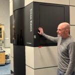
Gan Lab — 3D chromatin architecture and gene regulation: The Gan Lab investigates how the three-dimensional architecture of chromatin and nuclear molecular machines regulates gene expression inside native cellular environments. Using cryo-electron tomography, cryo-correlative light and electron microscopy, and advanced image reconstruction, the lab visualizes nuclear complexes in situ and connects chromatin organization to transcriptional control, genome regulation, and disease-relevant nuclear dysfunction.
Kenworthy Lab — Membrane microdomains as biosignaling organizing platforms: The Kenworthy Lab studies how membrane microdomains, including lipid rafts and caveolae, organize signaling, trafficking, and autophagy. By combining quantitative fluorescence microscopy, FRAP, single-particle tracking, and biochemical membrane assays, the lab defines how membrane heterogeneity, protein dynamics, and lipid–protein organization create functional platforms that maintain cellular homeostasis and become disrupted in disease.
Levental Lab — Lipid composition and membrane physical architecture: The Levental Lab investigates how lipid composition and dietary lipids shape the physical architecture of cellular membranes and thereby regulate signaling, metabolism, and cell physiology. Through lipidomics, membrane biophysics, model membrane systems, and quantitative imaging of membrane order, the lab connects lipid molecular structure to membrane organization, cellular regulation, and disease-relevant metabolic states.
Redemann Lab — Spindle architecture and genome stability: The Redemann Lab studies the molecular and physical principles that govern spindle architecture, chromosome movement, and faithful genome segregation. Using live-cell fluorescence microscopy, electron tomography, quantitative image analysis, and genetic perturbation, the lab reveals how cytoskeletal and chromosome-associated structures are assembled, remodeled, and coordinated to preserve genome stability during cell division.
Tamm Lab — Protein–lipid architecture of membrane remodeling and fusion: The Tamm Lab investigates how protein–lipid interactions drive membrane remodeling and fusion in processes such as viral entry, neurotransmitter release, and insulin secretion. Using structural biology, reconstituted membrane systems, biophysical fusion assays, and advanced microscopy, the lab defines how molecular architecture at membrane interfaces is converted into dynamic membrane function with major relevance to infection, neurobiology, and metabolism.
Collectively, these laboratories form a coherent ecosystem that integrates advanced imaging across scales—from molecular assemblies to whole cells and tissues—with quantitative biophysics and molecular engineering to understand how cells sense, organize, and respond to their environment, and how failures in these processes drive disease.