
Our Covers
Selected Publications
 Nature Communications, May 22, 2014
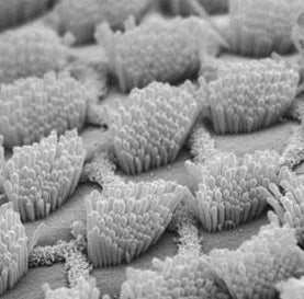
Hair cells pictured on the left are found in the auditory system of all
vertebrates and are important for sensing sounds. Thiede et al. analyze
gene expression patterns in chicken hair cells during embryonic development and find that retinoic acid signaling regulates the cellular organization in the inner ear. Read more at Nature.com.
 The Journal of Neuroscience, Volume 34, Number 5, January 29, 2014
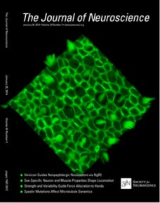
Responses to cell loss become restricted as the supporting cells in
mammalian vestibular organs grow thick junctional actin bands that
develop high stability
 The Journal of Neuroscience, Volume 32 , Number 19, May 9, 2012
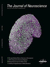
In vivo proliferative regeneration of balance hair cells in newborn mice
The Journal of Neuroscience, Volume 23, Number 10, August 17, 2011
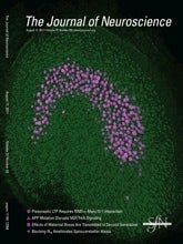
The postnatal accumulation of junctional E-Cadherin is inversely correlated with the
capacity for supporting cells to convert into sensory hair cells in mammalian balance
organs.
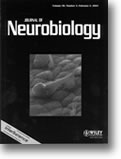 The Journal of Neurobiology, Volume 50, Number 2, February 5, 2002
A scanning electron micrograph of the surface of a bullfrog’s saccule that shows a hair
bundle and cuticular plate that have separated from the soma of a gentamicin-damaged
hair cell. Exposure to short-term, low-dose gentamicin causes loss of the sensory apparatus
from hair cells, which can survive and recover. Electron microscopy, time-lapse, and
multi-photon recordings were used to observe bundle separation, survival of bundleless
hair cells, and repair of damaged hair cell surfaces as described in Gale et. al.
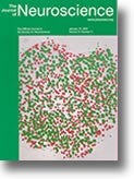 The Journal of Neuroscience, Volume 21, Number 2, January 15, 2001
Glial growth factor (GGF2) strongly stimulates S-phase entry in the early postnatal rat
utricle. DIC micrograph of a piece of rat utricular sensory epithelium composed only of
supporting and hair cells, and cultured for 72 hr in a medium containing GGF2. Nuclei of
cells that entered S-phase have incorporated BrdU (red). Fluorescent DAPI staining
revealed the nuclei that did not enter S-phase in the same piece of epithelium (green).
For details, see the article by Montcouquiol and Corwin in this issue (pages 570-580)
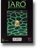 JARO Volume 1, Number 2, September 2000
Extramacular hair cell from the bullfrog saccule, labeled with HCS1, a specific hair cellantibody, in red. Green is phalloidin label. For a detailed description, see the paper by Gales, Meyers, and Corwin in this issue.